


Cleveland Medical Malpractice Attorneys Understand Errors That Lead to DVT and Pulmonary Embolism
What Exactly is a DVT?
DVT, deep vein thrombosis, is a blood clot that forms in a vein, generally deep in the body (away from the surface of the skin). DVTs generally form in the leg, calf or thigh, but can also form in the arms, pelvis, or abdomen. Complications of an untreated DVT fall into two main categories: 1) pulmonary embolism (blood clot in an artery of the lung); and 2) post-thrombotic syndrome. Complications of an untreated DVT can be devastating and life-threatening.
- Pulmonary Embolism (PE)
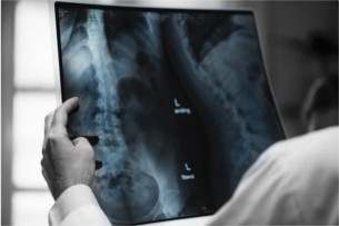
A Pulmonary Embolism is a blood clot in an artery in the lung. This blood clot causes blockage and prevents blood flow nourishment to a specific area of the lung. This can lead to death of lung tissue in this area. A PE can lead to low oxygen levels in the blood which then can lead to damage to other organs in the body, all of which need oxygen as nourishment. Pulmonary embolisms (the plural of “embolism” is also sometimes called “emboli”) are generally caused by DVT blood clots traveling to the lung and blocking an artery in the lung. A PE can cause shortness of breath, a dry cough, hemoptysis (coughing up blood), and sudden chest pain that worsens with deep inspiration (breathing in). When a large clot travels to the lung and blocks a large artery, more severe symptoms result, including intense pain and rapid death.
- Post-Thrombotic Syndrome (PTS)
More than one third of people who have DVTs go on to develop post-thrombotic syndrome (PTS). Symptoms of PTS include swelling, redness, ulcers and chronic leg pain. The primary cause of PTS is damage to the valves and walls of a vein due to the presence of a DVT. Prompt diagnosis and treatment of a DVT is necessary to prevent this damage from occurring. Once the valves and walls of a vein have been damaged, they cannot be repaired.
Risk Factors for Development of DVT
- An injury that damages the veins
- Overweight
- Family history of DVT
- Catheter placed in vein
- Hormone therapy or use of contraceptive pills
- Heavy smoking
- Flying or driving long distances - Sitting in the same position for long periods without any movement increases the risk of developing a DVT
- Prolonged bed rest, such as during hospital stay or paralysis
- Pregnancy – pregnant woman are 5 to 10 times more likely to develop a DVT. This increased risk continues until about six weeks after giving birth
- Hereditary blood-clotting disorders
- Inflammatory bowel disease
- Heart failure
- Surgeries, especially of the lower limbs – patients undergoing hip or knee replacement surgery are generally placed on anticoagulation medication for a period of time after surgery due to increased risk of developing a DVT
- Hypercoagulability (increased tendency for blood to clot) - This can either be related to recent surgery or trauma, presence of a malignancy (cancer) or an inherited hypercoagulable state (see below) .
Symptoms of DVT
- Swelling of foot, ankle, or leg, usually on one side
- Cramping of the affected leg that usually begins in the calf
- Severe pain in the foot and ankle
- Skin overlying the affected area is warmer than the skin overlying surrounding areas
- Bluish, reddish, or pale-colored skin over the affected area
- DVT can also be present without any symptoms
Evaluation for the Presence of a DVT
If a patient presents to a healthcare provider with any of the symptoms of a DVT, certain testing needs to be done to evaluate for the presence of a blood clot. First and foremost, a medical history and thorough physical exam should be performed. A medical history will be important to determine if any of the above risk factors are present. A simple, hands-on test for assessing for the possible presence of a DVT in the lower leg is to try to elicit the Homans Sign. The healthcare provider has the patient sit on the examination table with the affected leg out straight and the foot and ankle over the edge of the table. The examiner then quickly flexes the foot toward the shin. If there is a DVT, this flexion will cause pain in the calf muscles. While this procedure does not definitively diagnose DVT, a positive Homans Sign provides an indication for additional testing.
Another test that is often used as a screening test for a DVT is a D-dimer test, which is a simple blood test. A negative D-dimer is a good indication that there is no DVT. A positive D-dimer is not conclusive, as there are many other reasons why a D-dimer may be positive, but it a reason to continue looking for a possible DVT.
Other ways to look for a DVT include:
Ultrasound : This is the most commonly used test for diagnosing DVT. Ultrasounds use sound waves to create a picture of the vessels, and if a clot is present, an interruption of blood flow will be detected.
Venogram : Sometimes an ultrasound may be inconclusive and then a venogram may be done. This study involves dye being injected into the vein suspected of having a DVT. The dye makes the vein more visible and thus interruption of blood flow becomes more visible.
Recurring DVTs
If a patient develops recurrent DVTs and risk factors have been assessed and life-style changes made, then testing for inherited hypercoagulable states should be done. One common inherited state that causes recurrent DVTs is called the Factor V Leiden defect; others are antithrombin III deficiency, protein C or S deficiency and hyperhomocystinemia. All of these are conditions that cause the blood to clot more easily.
DVT Treatment
DVT is a serious medical condition. DVT treatment generally focuses on keeping the clot from getting larger, reducing the risk of developing more clots, and preventing the development of a PE due to a DVT breaking off and traveling to the lung. The three most common treatments are:
Medications : Medications to thin the blood such as heparin (while in the hospital), Lovenox (generally used as a bridge until blood testing indicates the blood is thin enough when starting Coumadin), and Coumadin (warfarin). There are also newer medications that thin the blood: Xarelto, Eliquis, and Pradaxa are examples. Although aspirin is known to be a blood-thinner, it is not the preferred choice for DVT treatment or for prevention of DVTs in someone at risk for developing a DVT. Aspirin is an alternative therapy for patients who cannot tolerate long-term use of blood thinners.
Compression Stockings : Wearing compression stockings on the legs helps reduce the incidence of developing clots by reducing swelling in the leg.
IVC (inferior vena cava) Filter : The major complication associated with DVT is a pulmonary embolism. This condition can be life-threatening. There are patients who are unable to take blood thinners. To decrease the risk of developing a PE from a DVT traveling up from the lower extremities, an IVC filter may be surgically implanted into the inferior vena cava (the largest vein carrying deoxygenated blood from the lower and middle body into the heart). The filter is designed to catch any blood clots traveling up towards the heart before those clots can be pushed on by the heart to the lungs. It is important to note that the effectiveness and safety of IVC are not very well-established, so placement of such filters generally are only recommended in limited, high-risk scenarios. Many of these devices can be removed and may be placed only for a short period of time if anticoagulation can be started in the future. IVC filter use does come with risks. Among other things, if multiple clots get caught in the filter, it can cause obstruction of the inferior vena cava, which can result in significant, additional problems. Another complication with the use of IVC filters is migration of the filter from where it was initially placed. It can migrate or travel to another part of the inferior vena cava or even to the heart. Filters can also fracture, causing pieces of the filter to travel to an to the heart or lungs, resulting in severe injury or death.
Medical Negligence
DVTs are generally quickly diagnosed and easily treated. Misdiagnosis or delay in treatment can lead to life-threatening conditions or lifelong leg pain. Delay in providing treatment, such as blood thinning medications or placement of IVC filters, in patients at high risk for developing blood clots may be considered medical negligence. Complications of an untreated DVT can be devastating and life-threatening.
If you or a loved have suffered injury or death due to complications from a DVT or PE, contact Cleveland’s experienced medical malpractice attorneys at The Eisen Law Firm for a free consultation today at 216-687-0900 or contact us online.